UCLA Unveils AI-Powered Autopsy Tissue-Staining Method Using Deep Learning
Advance eliminates cellular sample staining problems while expediting results
Ozcan Lab/UCLA
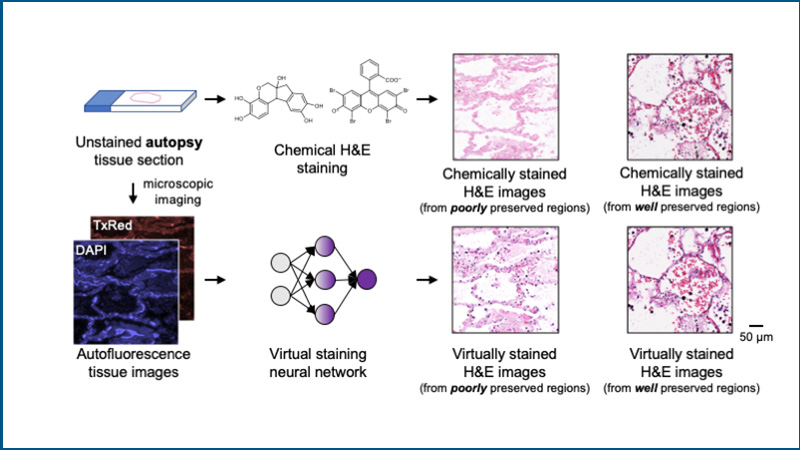
Images of label-free autopsy tissue sections from an AI-based virtual staining method compared to those generated by chemical staining
Autopsies play a central role in shedding light on disease pathophysiology and uncovering the causes of death. However, traditional autopsy chemical staining used for microscopic study of tissues and organs, or histology, suffers from delays in tissue preparation and preservation.
Now a UCLA research team led by Aydogan Ozcan, the Volgenau Chair for Engineering Innovation and an electrical and computer engineering professor at the UCLA Samueli School of Engineering, has developed a new technique to digitally process and stain autopsy tissue using artificial intelligence.
The current workflow for chemical staining is costly, time-consuming and labor-intensive. The method is also vulnerable to challenges that can arise from increased demand during a global health crisis, such as the COVID-19 pandemic.
By bypassing the need for physical staining of tissue, the virtual-staining approach is able to avoid delays in the preservation of biological tissue that result in decay due to autolysis — the process in which cells are “digested” by their own enzymes. Autolysis can result in changes to the tissue that interfere with the normal binding of staining dyes and biomolecules in the tissue, diminishing the quality of chemical staining and impacting the clarity and reliability of autopsy outcomes.
Published in Nature Communications, the UCLA study describes an AI-based staining approach that can process label-free autopsy tissue samples. The system uses a trained neural network to process and transform autofluorescence microscopic images of autopsy tissue sections to their hematoxylin and eosin (H&E)-stained counterparts. H&E staining is one of the primary stains used in histology to label different parts of a tissue section from an autopsy or biopsy. In addition to mitigating autolysis-induced decomposition observed in traditional chemical staining of autopsy samples, the approach is also faster, cheaper and easier.
“This is a green technology that eliminates tissue waste, and reduces manual labor, toxic reagents and waste water involved in the traditional staining process,” said Ozcan, who leads the Ozcan Research Group. “The method has great potential for widespread applications in histology.” Ozcan also holds faculty appointments in the Department of Bioengineering and the David Geffen School of Medicine at UCLA. He is the associate director of entrepreneurship, industry and academic exchange at the California NanoSystems Institute at UCLA.
“This is a green technology that eliminates tissue waste, and reduces manual labor, toxic reagents and waste water involved in the traditional staining process,” said Aydogan Ozcan.
The team demonstrated its technique in a series of blind tests, with pathologists from UCLA and other medical centers evaluating the staining quality of test images from autopsy tissue samples without knowing which images were virtually or chemically stained. The tests confirmed there was no observable difference in nuclear, cytoplasmic and extracellular details shown in the virtually and chemically stained H&E images obtained from well-preserved tissue regions. In fact, the virtual staining method clearly revealed much more detailed features, particularly in tissue samples severely affected by autolysis, such as COVID-19 samples, where notable delays in tissue preservation had occurred.
Funding for the research came from the National Science Foundation’s Biophotonics Program and the National Institutes of Health’s National Center for Interventional Biophotonic Technologies.
The research was completed in collaboration with Dr. Jonathan Zuckerman and Dr. Sepehr Hamidi from the Department of Pathology of the David Geffen School of Medicine at UCLA; Dr. Anatoly Urisman from the Department of Pathology at UC San Francisco; Dr. William Dean Wallace from the Department of Pathology at the USC Keck School of Medicine; and Dr. Tal Keidar Haran from the Department of Pathology at the Hadassah Hebrew University Medical Center in Israel.
Other authors of the paper are members of Ozcan Lab: UCLA Samueli postdoctoral scholar Dr. Nir Pillar, as well as Yuzhu Li, Jingxi Li, Tairan Liu, Di Wu, Songyu Sun, Guangdong Ma, Kevin de Haan, Luzhe Huang and Yijie Zhang.